Home
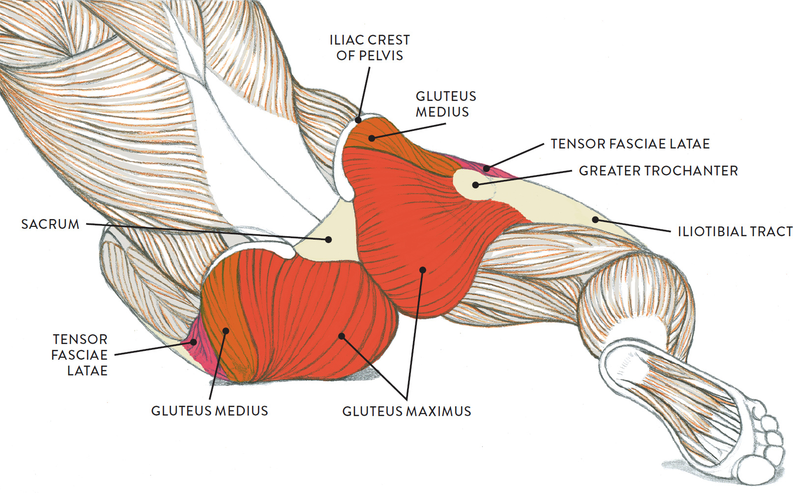
/ Diagram Of Hip.and Back.muscles, Remedial Massage for Hip Stabilisation : The hip muscle diagram below shows a number of the muscles we will be discussing in the next sections.
Diagram Of Hip.and Back.muscles, Remedial Massage for Hip Stabilisation : The hip muscle diagram below shows a number of the muscles we will be discussing in the next sections.
Diagram Of Hip.and Back.muscles, Remedial Massage for Hip Stabilisation : The hip muscle diagram below shows a number of the muscles we will be discussing in the next sections.. Lower back muscles below the shoulder blade. Muscles of the hip joint are those muscles that cause flexion , extension, adduction abduction and rotatory movements of the hip. The former two groups, superficial and intermediate, are referred to as the extrinsic back muscles. Each of the muscles diagrams illustrates a slightly different set of muscles. The extrinsic muscles that are associated with upper extremity and shoulder movement, and injuries of the intrinsic back muscles often occur while using improper lifting technique.
Back muscles are divided into two specific groups: Flexion of the trunk and thigh, lateral flexion of the trunk (excluding psoas major and minor only) innervation. Sit on the floor with your legs extended straight in front of you 2. Muscles of the upper limb (deltoid, biceps, forearms). Other muscles are small and cover much less space.
Sanguine and brown pastel pencils, white chalk on tone paper. from schoolbag.info Abducts and rotates thigh laterally, flexes knee at hip, originates at the anterior superior iliac spine and inserts on the medial surface of proximal tibia. Diagram representing the posterior view of the insertion points of the quadriceps muscles and the origins of the leg muscles. Dislocation of the hip joint. Back muscles anatomy lower back muscles anatomy human anatomy. Muscles of the deep back, adbominal wall, and pelv… Hip muscles and tendons march 19 2019 by luqman. Back muscles are divided into two specific groups: Back pain is the most common type of chronic is it any wonder that many consider the deadlift as the king of all exercises?
Related posts of muscles of the lower back and hip diagram muscle anatomy posterior.
Extension and lateral rotation at the hip. Muscles of the hip and lower limb. Lower back muscles below the shoulder blade. Some of these muscles are quite large and cover broad areas. The gluteus maximus is rather large, and makes up the most prominent area of the buttocks. Dislocation of the hip joint. Abducts and rotates thigh laterally, flexes knee at hip, originates at the anterior superior iliac spine and inserts on the medial surface of proximal tibia. The levator ani muscle along with a second muscle forms the pelvic floor. It joins the lower limb to the pelvic girdle. Back muscles are divided into two specific groups: The former two groups, superficial and intermediate, are referred to as the extrinsic back muscles. Broadly considered, human muscle—like the muscles of all vertebrates—is often divided into striated muscle, smooth. In human anatomy, the muscles of the hip joint are those muscles that cause movement in the hip.
The red lines show where the tendons attach the muscles to the bones. In human anatomy, the muscles of the hip joint are those muscles that cause movement in the hip. Abducts and rotates thigh laterally, flexes knee at hip, originates at the anterior superior iliac spine and inserts on the medial surface of proximal tibia. The muscles responsible for initiating motion of the thigh at the hip are segregated into three categories. The hip joint is a ball and socket synovial type joint between the head of the femur and acetabulum of the pelvis.
http://humananatomybody.info/anatomy-of-muscles-hip-and ... from i.pinimg.com It joins the lower limb to the pelvic girdle. Back muscles are divided into two specific groups: This is a table of skeletal muscles of the human anatomy. Deadlift muscles will include knee, hip, and back extensors, which primarily include the quads, glutes, and spinal erectors. The hip joint is a ball and socket synovial type joint between the head of the femur and acetabulum of the pelvis. Key facts about hip muscles. Most modern anatomists define 17 of these muscles, although some additional muscles may sometimes be considered. Some of these muscles are quite large and cover broad areas.
Dislocation of the hip joint.
Anatomy of the body hip muscles anatomy muscular system anatomy. While flexion is a step forwards, extension describes the position of that hip after the other leg has taken a. It joins the lower limb to the pelvic girdle. Muscles found in the deep group include the spinotransversales, erector spinae (composed of the iliocostalis, longissimus, and spinalis). All of these things can lead to long term back pain (and chronic complaining!). The hip muscle diagram below shows a number of the muscles we will be discussing in the next sections. There are anterior muscles diagrams and posterior muscles diagrams. You can protect the back muscles by bending from the hip and. Human muscle system, the muscles of the human body that work the skeletal system, that are under voluntary control, and that are concerned with movement, posture, and balance. There are around 650 skeletal muscles within the typical human body. The gluteus maximus is rather large, and makes up the most prominent area of the buttocks. Back muscles anatomy lower back muscles anatomy human anatomy. Muscles of the upper limb (deltoid, biceps, forearms).
In human anatomy, the muscles of the hip joint are those muscles that cause movement in the hip. Common hip and back pain causes include injury to muscles from overuse disc injurydegeneration or spinal stenosis. The image below shows the bones from the back side of the hand. Diagram of muscles and anatomy charts. Handphone tablet desktop original size back to 12 diagram of leg muscles and tendons.
Pin on Medicina from i.pinimg.com Extension and lateral rotation at the hip. Most modern anatomists define 17 of these muscles, although some additional muscles may sometimes be considered. This article looks at the anatomy of the back, including bones, muscles, and nerves. Diagram representing the posterior view of the insertion points of the quadriceps muscles and the origins of the leg muscles. Luckily you've found this page to help you. The levator ani muscle along with a second muscle forms the pelvic floor. It joins the lower limb to the pelvic girdle. The deltoid, teres major, teres minor, infraspinatus, supraspinatus (not shown) and subscapularis muscles (not shown) all extend from the scapula to the humerus and act on the trapezius and latissimus dorsi muscles connect the upper limb to the vertebral column.
Back pain is the most common type of chronic is it any wonder that many consider the deadlift as the king of all exercises?
This is a table of skeletal muscles of the human anatomy. Back pain is the most common type of chronic is it any wonder that many consider the deadlift as the king of all exercises? Anatomy of the body hip muscles anatomy muscular system anatomy. The image below shows the bones from the back side of the hand. Extension and lateral rotation at the hip. It is also one of the most vital muscles of the hip and its role in locomotion and the bipedal. Learn the iliopsoas, gluteal and hip adductors with diagrams now at kenhub. There are around 650 skeletal muscles within the typical human body. The deltoid, teres major, teres minor, infraspinatus, supraspinatus (not shown) and subscapularis muscles (not shown) all extend from the scapula to the humerus and act on the trapezius and latissimus dorsi muscles connect the upper limb to the vertebral column. Common hip and back pain causes include injury to muscles from overuse disc injurydegeneration or spinal stenosis. The hip muscle diagram below shows a number of the muscles we will be discussing in the next sections. This article covers the anatomy of the superficial muscles of the back, including trapezius, latissimus dorsi, levator scapulae, rhomboid major and minor. Iliacus, psoas major, and psoas minor main function:

